For centuries, seeing smaller meant building better lenses. Expansion microscopy took a different route. Instead of improving optics, it physically enlarges the specimen, embedding tissue in a water-absorbing polymer, the same kind of chemistry used in disposable nappies, and letting it swell. Structures that were separated by nanometres end up millimetres apart, comfortably resolvable with ordinary microscopes.
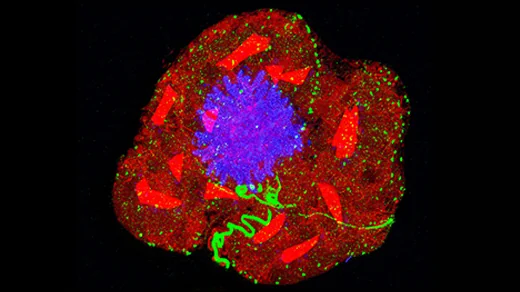
The approach has proved remarkably versatile. Because the swelling is uniform, distances are preserved and can be measured, turning a simple trick into a quantitative tool. Biologists have used it to trace the fine anatomy of synapses, to see how proteins organise themselves inside cells and to study microbes that are difficult to image any other way.
Expansion also plays well with existing techniques. It can be combined with fluorescent labels, with antibody staining and with tissue-clearing methods that make whole organs transparent. The result has been a genuine shift in what routine laboratories can see, and the technique keeps improving as researchers find ways to make the gels stronger and the expansion more accurate.